Improved Cardiac Shimusing Field Map Estimatefrom Multi-echo Dixon Method


Peter Kellman1, Saurabh Shah2, Diego Hernando3,
Sven Zuehlsdorff2, Andreas Greiser4, Renate Jerecic2
1National Institutes of Health, Bethesda, MD2Siemens Medical Solutions, Chicago, IL
3University of Illinois, Urbana, IL
4Siemens Medical Solutions, Erlangen, Germany

Motivation:for improved cardiac shim
•B0-field inhomogeneity affects images …
•GRE-EPI artifacts
•SSFP dark banding artifacts
•Chemical shift fat suppression
•T2* losses
•Rapid field variation across the heart
•caused by tissue-air interface & veins

Approach:Joint fat/water/field map estimates
•multi-echo GRE acquisition
•VARPRO solution for fat and water components &fieldmap estimate
•parallel imaging reconstruction
•real-time, free-breathing, non-ECG gated 2D
• multi-slice acquisition, acquiring the volume in ≈ 7s

water & fat separation:multi-echo Non-linear Least Squares methods
multi-echo
dataset


VARPRO
Signal model:
ML cost function:
VARPRO formulation:

Sequence:real-time, multi-echo GRE
Typical parameters:
matrix size:64x48
parallel imaging:rate 2 (GRAPPA)
number of echoes:6 (monopolar readout)
bandwidth:900 Hz/pixel
TE (TR):1.2, 2.7, 4.3, 5.8, 7.3, 8.9 ms (10.25 ms)
duration:246 ms/slice
spatial resolution:6.25 x 6.25 mm2 (8 mm thick with no gap)
# slices:20 slices typical (covering 160 mm)
1 2 3 M=48
N-echoes per PE line
Acquire auto-calibration data for parallel imaging
1 2 3 4 5 6 7 8 9 10 11 12 13 14 15 16 17 18 19 20
Slice
approx 7 sec

Volume coverage2d multi-slice


Improved cardiac shim

SSFP
cine
fieldmap
before
after
before
after
0
20
40
60
80
100
120
140
-300
-200
-100
0
100
profile position (mm)
frequency (Hz)

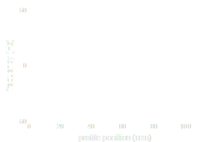
Real-time fieldmap estimatesfree breathing, non-triggered
fieldmap
(±200 Hz)
Fieldmap profiles across heartfor 50 repetitions using shimsequence.

magnitude
std = 4 Hz

Summary
•Approach to field map estimation
• multi-echo fat/water separation imaging
• low spatial resolution fieldmap
• non-triggered, free-breathing acquisition
• real-time with parallel imaging
•2d-multislice volume coverage
•Performance
• reliable and fast (<7.5 s / volume)
• relatively insensitive to cardio-respiratory phase